Imaging
The Madison County Memorial Hospital Imaging Services Department (also called Radiology) provides a team of skilled professionals who are available to administer diagnostic radiological procedures as prescribed by your physician. All images are read by a board-certified radiologist. The majority of our technologists are cross-trained in more than one skill, and many have dual certifications. All are registered by the American Registry of Radiologic Technologists and are licensed with the State of Florida’s Bureau of Radiation Control. All diagnostic equipment is digital. This makes for a more robust health record and allows your physician to access your radiology image from anywhere in the world. To explore the various imaging/radiology services available, please click on the tabs below.
Mammography
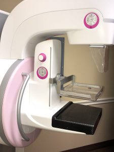
Madison County Memorial Hospital  provides its patients with the highest quality of care in the prevention and early detection of breast cancer. Mammography uses low-dose x-rays to image the breast tissue for nodules, cysts, or calcifications.
Mammography allows the radiologist to view the X-ray image closely, zeroing in on suspicious or concerning areas and enabling them to make decisions about additional images. Your mammogram image is run through a special computer that detects breast nodes too small for the human eye to detect. The Computer Aided Detection (CAD) image, and a detailed report, is given to the radiologist which helps insure that you get the care you need.
Ultrasound
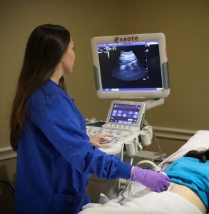
 This device uses ultrasonic frequency waves that pass through the body and painlessly bounce off the internal organs, producing an image electronically. Ultrasound is used to detect changes in the appearance of organs, tissues, and vessels. It is also used to detect abnormal masses, such as tumors and cysts.
It is used to help diagnose the causes of pain, swelling and infection in the body’s internal organs and to examine a baby in pregnant women and the brain and hips in infants. It’s also used to help guide biopsies, diagnose heart conditions, and assess damage after a heart attack. Ultrasound is safe, noninvasive, and does not use ionizing radiation.
This procedure requires little to no special preparation. Your doctor instructs you on how to prepare, including whether you should refrain from eating or drinking beforehand. It is best to leave jewelry at home and wear loose, comfortable clothing. Depending on the procedure, you may be asked to wear a gown.
Computerized Axial Tomography (CAT or CT Scan)
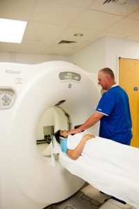
 CT scan (or CAT scan as it is sometimes called) stands for Computerized Axial Tomography. CT scans can detect bone and joint problems. For example, complex bone fractures and tumors. They show internal injuries and bleeding, such as those injuries from a car accident. If you have a condition like cancer, heart disease, emphysema, or liver masses, CT scans spot these types of abnormalities and helps doctors see changes along the way.
The CT scanner can do head-to-toe imaging in minutes by taking a lot of pictures of your body from different angles. These pictures are fed into a computer. The computer puts them together to give a series of cross sections or ‘slices’ through the part of the body being scanned. A very detailed picture of the inside of the body can be built up in this way.
Computed Radiology (CR)
 General Diagnostic Radiology: consists of a variety of radiological examinations that image bony structures, soft tissue and various internal organs.
The Computed Radiography (CR) system quickly produces and manages digital images. The CR manages data from throughout the hospital and quickly produces high-quality digital images. Specifically designed, CR ensures images and reports are available at the right place and the right time.
The digital radiography system is designed to improve workflow while being patient-and-clinical friendly. Multiple exams can be conducted without moving the patient. The flat panel technology means the equipment can be brought to the patient. For individuals in a wheelchair or on a gurney, this means increased comfort and faster exams. For clinicians, this intuitive technology means fewer retakes, faster examinations, and integration with the digital environment.
PAC Systems
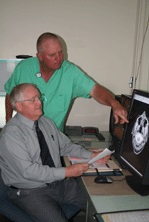
 PAC System stands for picture archiving and communication system. This system provides imaging technology to store your images in a save, economical and convenient way. Carrying copies of our radiology films and CD’s to your physician is no longer necessary. Because all of your images are digital, your physician can see your X-ray simply by pulling it up on his or her computer screen. This service helps our clinicians, your physician and you gain access to you images to see and share files and make informed decisions about your care plan. The privacy of your images are protected by a private code that your physician enters.
Radiology Information System (RIS)
 The Radiology Information System (RIS) streamlines information into a central resource. Radiology registration, scheduling, tracking, image management and transcription are housed in one electronic system. This central resource gives clinicians access to the information they need to make a diagnosis and to establish a care plan for you. Patient information, including historical information, is available with the click of a mouse. The result: faster, more accurate information.
